

Material: medical pure titanium
Thickness: 1.4mm
Product specification
|
Item No. |
Specification |
|
|
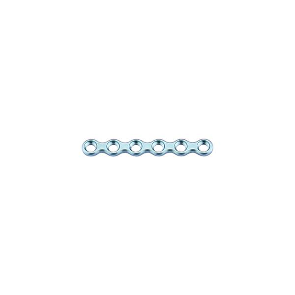
10.01.04.06011235 |
6 holes |
35mm |
|
10.01.04.08011200 |
8 holes |
47mm |
|
10.01.04.12011200 |
12 holes |
71mm |
|
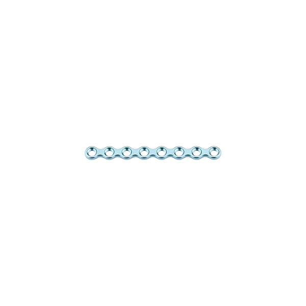
10.01.04.16011200 |
16 holes |
95mm |
Features & Benefits:
• locking maxillofacial micro and mini plate can be used reversibly
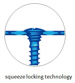
• locking mechanism: squeeze locking technology
• one hole select two kinds of screw: locking and non-locking are all available, probabilize the free collocation of plates and screws, meet the demand of clinical indications better and more extensive indication
• bone plate adopt special customized German ZAPP pure titanium as raw material, with good biocompatibilty and more uniform grain size distribution.Don't affect MRI/CT examination
• bone plate surface adopt anodizing technology, can enhance surface hardness and abrasive resistance
Matching screw:
φ2.0mm self-tapping screw
φ2.0mm locking screw
Matching instrument:
medical drill bit φ1.6*20*78mm
cross head screw driver: SW0.5*2.8*95mm
straight quick coupling handle
Orthopedic surgery or orthopedics, is one branch of surgery. Orthopedics care about musculoskeletal system. Both surgical and nonsurgical means are used by orthopedic surgeons to treat musculoskeletal trauma, spine diseases, sports injuries, degenerative diseases, infections, tumors, and congenital disorders.
Top 25 most common procedures performed by orthopedic surgeons in order are: knee arthroscopy and meniscectomy, shoulder arthroscopy and decompression, carpal tunnel release, knee arthroscopy and chondroplasty, removal of support implant, knee arthroscopy and anterior cruciate ligament reconstruction, knee replacement, repair of femoral neck fracture, repair of trochanteric fracture, debridement of skin / muscle / bone / fracture, knee arthroscopy repair of both menisci, hip replacement, shoulder arthroscopy / distal clavicle excision, repair of rotator cuff tendon, repair fracture of radius (bone) / ulna, laminectomy, repair of ankle fracture (bimalleolar type), shoulder arthroscopy and debridement, lumbar spinal fusion, repair fracture of the distal part of radius, low back intervertebral disc surgery, incise finger tendon sheath, repair of ankle fracture (fibula), repair of femoral shaft fracture, repair of trochanteric fracture.
maxillofacial trauma in children as well as adults common come from sports injuries, falls, assaults, vehicle crashes, blunt assaults, blows from fists or objects. Animal attack, gunshots, blasts and other wartime injuries can also cause facial bone fracture. Vehicular trauma is one of the leading causes of facial injuries in city lives. Trauma commonly occurs when the face strikes a part of the vehicle's interior, such as the steering wheel. In addition, airbags can cause corneal abrasions and lacerations to the face when they deploy.
Facial bone injuries can be roughly divided intoinclude the nasal bone, the maxilla, and the mandible. The mandible may be fractured at its symphysis, body, angle, ramus, and condyle. The cheekbone and the frontal bone are other places for fractures. Fractures may also occur in the bones of the palate and those that come together to form the orbit of the eye.
At the beginning of the 20th century, René Le Fort mapped typical locations for facial fractures; these are now known as Le Fort I, II, and III fractures (right). Le Fort I fractures, also called Guérin or horizontal maxillary fractures, involve the maxilla, separating it from the palate. Le Fort II fractures, also called pyramidal fractures of the maxilla, cross the nasal bones and the orbital rim. Le Fort III fractures, also called craniofacial disjunction and transverse facial fractures, cross the front of the maxilla and involve the lacrimal bone, the lamina papyracea, and the orbital floor, and often involve the ethmoid bone, are the most serious. Le Fort fractures, which account for 10–20% of facial fractures, are often associated with other serious injuries.
Surgical treatments are adopted to repair maxillofacial bone fracture, aim to repair the face's natural bony architecture and to leave as little apparent trace of the injury as possible. Bone injuries may be treated with pure titanium plates and titanium alloy screws. Resorbable materials are another choice available.
maxillofacial trauma rarely bring a threat to life, but it is often associated with dangerous injuries, airway blockage and other life-threatening complications. The airway can be blocked due to bleeding, swelling of surrounding tissues, or damage to structures. Burns to the face can cause swelling of tissues and thereby lead to airway blockage. Combinations of nasal, maxillary, and mandibular fractures can influence with the airway. It is necessary to monitor the airway regularly, because airway problems can occur late after the initial injury.
bones need to be put back into their proper places as soon as possible, because nerves and muscles may be trapped by broken bones. Orbital floor fracture or medial orbital wall bone fracture of the eye can entrap the medial rectus or inferior rectus muscles.
In facial wounds, tear ducts and nerves of the face may be damaged. Fractures of the frontal bone can interfere with the drainage of the frontal sinus and can cause sinusitis. Infection is another potential complication.
-
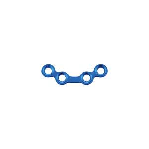
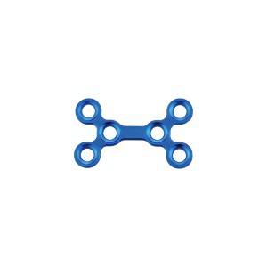
locking maxillofacial mini arc bridge plate
-
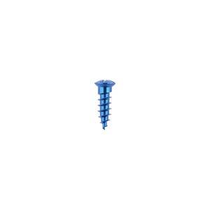
φ2.0mm self-drilling screw
-
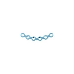
locking maxillofacial micro straight bridge plate
-
maxillofacial trauma mini arc plate
-
orthognathic 0.8 genioplasty plate
-
locking maxillofacial mini double Y plate