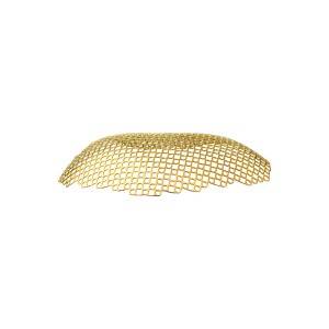
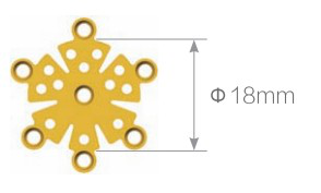
Material: medical pure titanium
Product specification
|
Thickness |
Item No. |
Specification |
|
0.6mm |
12.30.4010.181806 |
Non-anodized |
|
12.30.4110.181806 |
Anodized |
Features & Benefits:
• No iron atom, no magnetization in magnetic field. No effect to ×-ray, CT and MRI after operation.
• Stable chemical properties, excellent biocompatibility and corrosion resistance.
• Light and high hardness. Sustained protect brain issue.
• Fibroblast can grow into the mesh holes after operation, to make the titanium mesh and tissue integrated. Ideal intracranial repair material!
Matching screw:
φ1.5mm self-drilling screw
φ2.0mm self-drilling screw
Matching instrument:
cross head screw driver: SW0.5*2.8*75mm
straight quick coupling handle
cable cutter (mesh scissors)
mesh moulding pliers
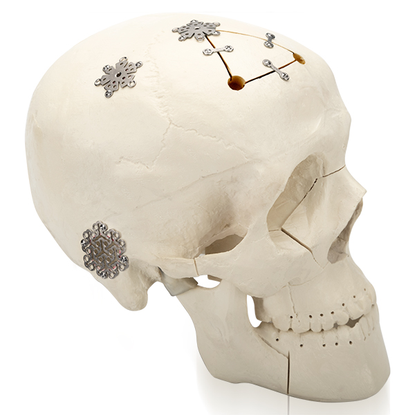
cranial (from Greek κρανίον 'skull') or cephalic (from Greek κεφαλή 'head') describes how close something is to the head of an organism.
The defect of skull is partly caused by open craniocerebral trauma or firearm penetrating injury, and partly caused by surgical decompression, skull lesions and puncture damage caused by skull resection.There are the following etiologies: 1. Open craniocerebral trauma or firearm puncture injury.2. After reamation for comminuted or depressed skull fractures that cannot be reduced.3. The severe traumatic brain injury or other types of craniocerebral surgery because of illness need to bone disc decompression.4. Growing skull fracture in children.5. Cranial osteomyelitis and other lesions of the skull itself caused by puncture skull destruction or surgical resection of skull lesions.
Clinical manifestations: 1. No symptoms.Skull defects smaller than 3cm and those located below the temporal and occipital muscles are usually asymptomatic.2. Skull defect syndrome.Headache, dizziness, nausea, loss of limb strength, chills, tremors, inattention and other mental symptoms caused by a large skull defect.3. Encephalocele and neurolocational signs.In the early stage of skull defect, severe brain edema, the dural of brain tissue and the formation of fungoidal bulge at the skull defect, which was embedded at the bone margin, caused local ischemic necrosis and caused a series of neurological localization symptoms and signs.4. Bone sclerosis.The area of skull defect caused by growth fracture in children expands continuously, and the bone sclerosis around the defect forms.
Cranial repair is the main treatment strategy for skull defect.Indications for operation: 1. Cranial defect diameter BBB 0 3cm.2. The diameter of the skull defect is less than 3cm, but it is located in the part affecting aesthetics.3. Pressure on the defect can induce epilepsy and meninge-brain scar formation accompanied by epilepsy.4. Skull defect syndrome caused by skull defect causes mental burden, affects work and life, and has the need for repair.Surgical contraindications: 1. Intracranial or incision infection has been cured for less than half a year.2. Patients whose symptoms of increased intracranial pressure have not been effectively controlled.3. Severe neurological dysfunction (KPS <60) or poor prognosis.4. The scalp is thin due to extensive skin scar, and the repair may cause poor wound healing or scalp necrosis.Timing of operation and basic conditions: 1. The intracranial pressure has been effectively controlled and stabilized.2. The wound healed completely without infection.3. In the past, 3 ~ 6 months of repair after the first operation was recommended, but now 6 ~ 8 weeks after the first operation is recommended.The reimplantation of autologous bone flap buried within 2 months is appropriate, and the traction reduction method of subcapate aponeurosis buried should not exceed 2 weeks.4. Cranial repair is not recommended under 5 years old because the head and tail grow rapidly;5 ~ 10 years old can be repaired, and overburden repair should be adopted, and the repair material should be 0.5cm beyond the bone margin.After 15 years of age, skull repair is the same as in adults.Commonly used repair materials: high polymer material, organic glass, bone cement, silica, titanium plate), allograft bone material use less (has), allograft material (like the kind of allograft decalcified, degreasing and other processing made of bone matrix gelatin), autologous materials (ribs, shoulder blades, skull, etc.), new materials, porous high density polyethylene, EH composite artificial bone), the current in the shape of 3 d reconstruction of titanium plate is most commonly used.
-
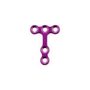
maxillofacial trauma micro T plate
-
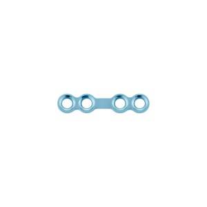
locking maxillofacial mini straight bridge plate
-
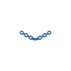
locking maxillofacial mini 120° arc plate
-
anatomical titanium mesh-2D round hole
-
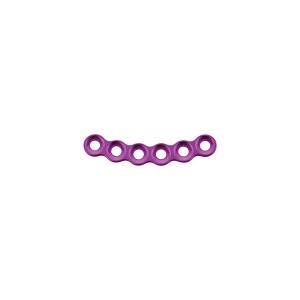
locking maxillofacial micro arc plate
-
maxillofacial trauma mini arc plate